
Microvascular Damage - With and without SARS-CoV-2
Journey into the Microvascular Abyss

In my journey towards understanding the complexity (heh) of SARS-CoV-2's impact on our physiology, it led me towards researching many scientific and medical fields, conditions, and experiencing many of the effects I have covered myself. Among the most under-researched, with the most long-term consequences of a chimeric infection lies the issue of micro-clotting, or more broadly and perhaps more accurately, vast and significant damage and dysfunction within your microvasculature.
Before going through the paper and its significance, I will leave some suggestions on how to address what is to be discussed. Most supplements cited here have articles explaining their use in my Substack and by other authors, too.
Resveratrol and Curcumin both inhibit necroptosis.
Olive Leaf Extract is one of the most powerful supplements for endothelial health, recovery, and complement modulation, too.
Quercetin and a few other compounds can activate your antioxidant system, and scavenge the content released from RBCs
Omega-3 will help improve RBC and other cell membranes
Lastly, Gingko biloba. It acts directly in the endothelial, it regulates blood flow, and it is one of the few supplements that can act in the capillaries
Aspirin, Activated Charcoal, but especially BHB, an exogenous Ketone to deal with HMGB1, and a lot of what is discussed here
Microvascular damage is a significant contributor to many diseases, including post-septic conditions, viral reactivation triggered by excessive inflammation, and, of particular interest, neurodegeneration. Microvascular dysfunction or damage is one of the driving forces of Alzheimer’s Disease. This damage can also affect organs themselves, and over time develop specific diseases, such as liver or kidney diseases.


There is a significant interplay between shifts in your immune cells, inflammation, and microvascular damage and dysfunction. Neutrophils, macrophages, lymphocytes, your T-cells and their T Helper cells, a shift in those, and microvascular damage may occur, among a myriad of other effects. Thus, today, we cover an underappreciated cell group, but among the most important cells we have, and how it contributes to such damage. Red Blood Cells.
Thanks to my reader Dee for getting this paper for me (paywalling in 2025 is outrageous).
Ischaemic endothelial necroptosis induces haemolysis and COVID-19 angiopathy
SARS-CoV-2 has a predilection, a preference towards infecting ACE2-rich cells, but it may also bypasses this primary method and use other receptors, by both directly infection and indirectly damaging the endothelial cells (the cells that make up the “lining” of your veins) it can induce significant long-term sequelae and contribute to heart disease, stroke, and Long Covid.
As we covered microthrombi formation as a byproduct of infection, we have seen it is often formed either by proteins, small fragments (platelets are small fragments), or sometimes a misshapen collection of these with fibrin and the infamous amyloid. Amyloid-based thrombi, small or bigger alike, are all resistant to being dissolved, which makes them pernicious and lasting. Rather troublesome that when faced with persistent microthrombi, medical anticoagulation therapy does little to improve symptoms.
As usual with scientific research, solving new pieces of a complex puzzle, you need to physically observe the changes you hypothesised at first. The authors developed a multimodal imaging method to quantitatively analyze Endothelial Cells (ECs from here on) from autopsy patients, with samples coming from lungs, heart, kidney, and liver (usually the main organs hit by systemic failure).
Quantitatively means the authors actively measure the levels of specific markers, and score changes in the cells, multimodal imaging means using multiple imaging methods to observe structural changes in the tissues. They analysed ECs from more than 1.000 vessels throughout the arterial, venous, and capillary networks from the specific type of patients they selected, those who suffered from multi-organ dysfunction.

Widespread damage to ECs was apparent throughout the microcirculation of all chosen organs, evidenced by physical (morphological) changes that are typical of cell death, and the extent of EC death correlated with the degree of tissue injury, and so did markers for such, VE-cadherin, PECAM-1, and glycocalyx, this last one of a hidden yet very significant importance.
By using microscopy imaging to look into the autopsy tissues, they observed an amorphous, proteinaceous acellular material in the places where Endothelial Cell death occured, such as in the microvasculature of the heart (30%), kidney (29%), and liver (27%), but significant less in the lungs (5%).
Using a staining material, the authors were able to observe that said amorphous material was primarily derived from Red Blood Cells (RBCs), and it was not recognized by tests that react to fibrin and platelets, or DNA. Using electron microscopy, they were able to confirm these were haemolysed Red Blood Cell membranes, meaning when RBCs suffer damage and rupture, their membranes stick together, forming these “clumps” that are deposited in the inner lining of the vasculature and between adjacent RBCs.
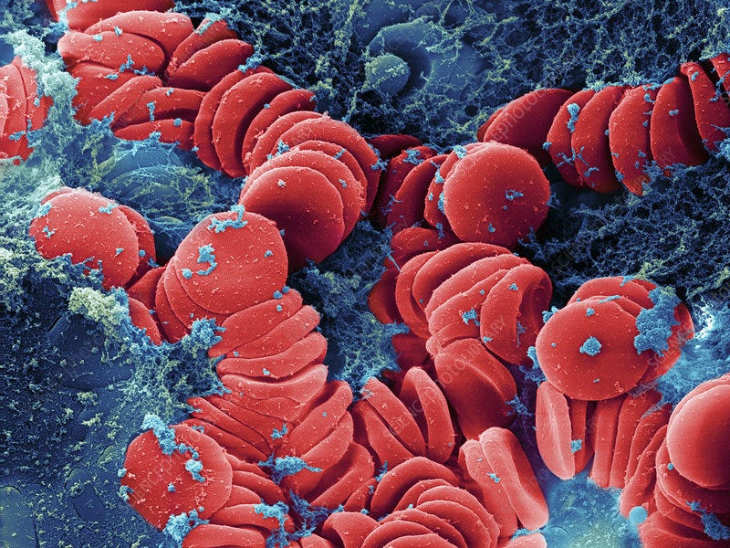
These RBC aggregates had a distinct shape compared to fibrin polymers. They then compared tissue samples from non-COVID-19 ARDS (acute respiratory distress syndrome), which usually don’t suffer from multisystem (many organs) involvement. In patients with Covid, microvascular RBC haemolysis (rupture and destruction of red blood cells) was prominent in the kidneys and liver, yet in non-COVID, the same was minimal. Minimal lung involvement in both.
Fragmented RBCs, called schistocytes, which are misshapen, occur in 70% of hospitalized Covid patients, and it is argued that it is the secondary cause for microvascular fibrin deposition. This occurs in DIC, Disseminated Intravascular Coagulation, a condition I wrote about a few times, and here they found minimal evidence for it, but found a strong correlation between the RBC clumps and Endothelial Cell death.
To preface the next sections, I will try to summarize in a shorter and less complex manner because we have more to cover beyond this paper, but directly linked to its contents.
RBC destruction induced by poor organ blood flow
To understand the mechanism that causes the aforementioned systemic damage, and determine if the RBC destruction was specific to SARS-CoV-2 infection or a consequence of ischemia (poor blood flow), they first analyzed the microvasculature from human tissue with other ischaemic condition, such as AMI (Acute Myocardial Infarction), stroke, gut ischaemia, septic and cardiogenic shock that never had Covid.
Non-Covid samples showed extensive microvascular RBC destruction (haemolysis) and EC death, with deposition patterns almost identical to what was seen in SARS-CoV-2 samples. Using mouse models, SARS-CoV-2 caused limited organ damage and only mild haemolysis, not matching the severe injury pattern seen in the lungs or brain. But in the mouse model for cardiac and gut ischaemia, extensive RBC haemolysis occurred in the regions with poor, low blood flow, and this RBC destruction was associated with endothelial cell death.
Haemolysis, the process by which Red Blood Cells die, is linked to the death of endothelial cells, which is caused by poor blood flow to the issue. Their next test sought to understand which type of cell death actually contributes to this mechanism, and they found something remarkable.
RBC haemolysis and its consequential clumping happened at specific sites in tissue where the endothelial cells went through necroptosis during ischaemia, necroptosis is a form of cell death that has specific pathways, and it is somewhat chaotic, inducing more inflammation. On the other hand, platelet and fibrin clots were formed primarily by apoptotic ECs during the restoration of blood flow, called reperfusion, apoptosis is the more common, less chaotic form of cell death.

Next, they sought to understand the molecular mechanisms by which this occur, which is necessary for proper targeting and treatment. The stop and restoration of blood flow (called Ischaemia-Reperfusion injury, IR injury) is known to induce necroptosis, but necroptosis itself has yet to be connected with RBC haemolysis.
Mixed Lineage Kinase domain-Like (MLKL) is the main mediator of the necroptotic pathway, and when it is activated, it forms pores in cell membranes, leading to cell lysis (the cell opening up and leaking its contents). When necrotic cells die, they are known to trigger the immune system, which can pierce holes in cell membranes via the Membrane Attack Complex (MAC), which requires a protein from the complement system, C9.
Using genetically modified mice lacking either MLKL in their Endothelial Cells or C9, they performed a gut IR injury and found that mice lacking MLKL in their Endothelial Cells had significantly less RBC haemolysis. Mice lacking C9 had markedly less haemolysis.
Imaging showed complement system components like C5b-9 and C9 deposited directly onto the lysed RBCs and specifically co-localised with the necroptotic ECs that had MLKL, not the apoptotic ones. This demonstrates ischaemia leads to EC necroptosis, which triggers complement activation, which in turn lyses (ruptures) RBCs, especially the ones nearby.
Similar C9 deposition on lysed RBCs and necrotic ECs was also evident in the microvasculature of the gut, heart, and kidney tissue samples from humans with and without Covid.
Microplugs for Microleaks
They then examined microvascular bleeding in the gut after stop and restoration of blood flow in mice, comparing bleeding in WT mice (they have the genes and proteins to go through haemolysis), MLKL and C8 Knockout mice (with reduced haemolysis, because they lack the genes/proteins). Surprisingly they found interstitial bleeding (inside organs/around cells) was more severe in areas where RBC haemolysis was low or absent.
Mice without MLKL and C9 showed a marked increase in microvascular bleeding compared to WT mice. Inhibiting platelets or coagulation also caused bleeding, but with distinct patterns from the other mice. Real-time imaging showed that during low-flow conditions such as ischaemia, RBCs would stick to the surface of dying Endothelial Cells, elongate and fragment, and their membrane remnants would deposit onto the vessel wall, and these fragments served as docking sites for RBCs in free flow, triggering a feedback loop.
These deposited layers of haemolysed RBCs membranes formed a continuous “endothelial vascular seal” (or endo-seal for short) lining the vessel wall. This seal structure was also seen in human ischaemic and Covid tissues. Quantitative analysis showed a strong inverse correlation. Where this endo-seal formed, bleeding was prevented. Haemolysed RBC membranes acted as a rapid, localised endovascular sealant on damaged endothelium.

So, how does extensive microvascular becomes a problem, sometimes inducing systemic microvascular bleeding and damage, inducing “macro” (your normal) vasculature damage, and clotting occur if the Red Blood Cells are forming microplugs ? Well, the adhesive and propensity of these RBC membranes to stick together has a downside. The RBC membranes only clump during very low shear rates, meaning, only when the blood flow is low enough (less than 25 s−1), above this rate, primarily platelet deposition occurred, and platelets and RBCs aggregates formed distinct “shapes”.
The mechanism of the paper is as it follows, during ischaemia, or low/insufficient blood flow, Endothelial Cells start dying, and mostly from a form of cell death called necroptosis. As they die, they release signals and inflammatory proteins, starting a chain reaction and activating the complement system nearby. This complement activation then ruptures (lyses) nearby Red Blood Cells.
These haemolysed RBC membranes are extremely sticky, and they deposit in the vessel lining where it is damaged, forming endothelial micro-plugs that prevent micro-leaks, preventing bleeding. But this stickiness has a cost, it starts a general clumping of membranes and intact RBCs, and in a state of low flow, these create microvascular obstruction, leading to tissue injury, initiating a feedback loop leading to broader organ dysfunction. We are faced with another double-edged sword.
The Things Hidden (or how amplification happens)

This is where things get a bit complicated. The mechanism described here is one independent of viral infection for the most part, microvascular dysfunction that can lead to systemic vascular damage can occur mostly without infection, by the simple yet significant change in blood flow, which was the focus of the paper.
Here we focus on the hidden trends, expanding on their great work. SARS-CoV-2 is known to directly infect erythroid precursor cells, which are derived from Stem Cells (covered in great detail above, with a lot more further reading inside). A mild infection can cause months-long impact on Red Blood Cells, with special attention to capillary blood flow impairment. The evidence is significant, and I may now go down this route. The Spike Protein itself causes RBCs to aggregate (in a different manner than describe in this paper).
The viral infection has numerous ways of directly and indirectly inducing EC death, not just by directly infecting them, but by a multifaceted mechanism.
Galectin-3 is known to interact with a huge number of receptors, and regulate immune responses, often enhancing inflammatory ones, both dying ECs and haemolysed RBC membranes have altered surfaces, allowing Gal-3 to interact with them and amplify, literally, each step of the pathway the authors found.
Th17, a subset of T helper cells of major importance, known to play a role at every stage of the infection, from protection to lasting damage, is known to play a central role in inflammatory conditions, autoimmune diseases. Th17 alone can activate Endothelial Cells and make the environment (especially the brain) more permeable, leaky. IL-17, its main cytokine, is also highly inflammatory.
Staphylococcal Enterotoxin B is a bacterial superantigen that causes severe, incredibly rapid activation of T-cells and can induce cytokine storm, and the leading cause of Toxic Shock Syndrome. SEB can cause profound and systemic, widespread hypoperfusion, meaning low blood flow, and can easily lead to widespread micro and macrovascular pathology. The pathway described by the authors is precisely one observed during severe superantigenic reactions.
Endotoxin, or LPS. One of the most well-studied and well-known bacterial toxins in the world, it primarily acts via the Toll-like Receptor 4, it doesn’t produce as drastic as a reaction as SEB, but it is one of the most potent and present toxins around and inside each of us. LPS can directly activate ECs via TLR4, and via Galectin-3, inducing inflammation and making them susceptible to death. Secondary bacterial infections after a mild SARS-CoV-2 infection are now so widespread, you can get antibiotics prescribed while sick with it.
This is one of the main drivers of sepsis and septic shock, both conditions the poster child for microvascular disease, especially lasting damage to the microvasculature. This is also one of the most (unknowingly by the authors and researchers) most studied Spike Protein damage mechanisms, because many of the commercially available Spike Protein comes contaminated with Endotoxin. Which to me is evidence for the main pathway, rather than a downside.
Lastly, Galectin-9. Omicron’s most pertinent and undertracked protein, also abundant when you are recently vaccinated or infected, high levels in any severe case. Galectin-9 role here is indirect. Gal-9 is often release during cellular stress, inflammatory responses or cell death, and while it can induce cell death, it is not its main role.
Galectin-9 will perpetuate immune dysfunction, as the body attempts very hard to maintain multiple immunological and inflammatory triggers in place, in turn, it contributes to EC death and many forms of vascular dysfunction. No other place its an impact in Red Blood Cells, low-grade inflammation, and immune dysfunction better than in this paper (I intend to write a whole article on this paper and Gal-9 alone).
Diverse immunological dysregulation, chronic inflammation, and impaired erythropoiesis in long COVID patients with chronic fatigue syndrome
Here, Gal-9 plays a central role in systemic immunological, low-grade inflammatory dysfunction, impairing erythropoiesis, or the production of new Red Blood Cells.
Lastly, but not last, a bridge to connect all of this and more. HMGB1. One of the most versatile proteins in the human body, possessing so many functions, it alone has 400 papers in its folder in my files. HMGB1 is passively released from any cells undergoing stress and death. Whenever a cellular membrane is damaged or ruptured, HMGB1 is released.
HMGB1 serves as a potent signal, and while it is not the cause of the initial loss of proper blood flow, it will sensitise other ECs to being damaged and die, and alter blood flow itself, amplifying the initial loop. HMGB1 released from dead or dying cells will bind to other dying or nearby cells in the area without proper blood flow, creating a feedback loop.
It plays a central role in vascular and microvascular diseases, and an intricate role with Red Blood Cells. We now not only possess insight into a novel mechanism on how RBCs can induce microvascular damage, but also a insight into the complexity (heh) of how microvascular leaks can cascade into systemic problems, and we have our first step towards a systemic understanding on how and why SARS-CoV-2 can impact the microvasculature and microstructures of the body.
Consider becoming a paid subscriber, and if you are one, thank you !

I hope all my readers are well. Most of the interesting papers now are highly complex and I need to layer them in a way that you can understand without remembering other 30 articles I wrote.
The next article should take much shorter and another of my forecasts, proven right. Sadly.
Nice to see the reference to contaminated commercial Covid19 Spike protein.
Enterotoxin and Endotoxin work in synergy to destroy embryos.
https://geoffpain.substack.com/p/enterotoxin-synergy-with-endotoxin