
Hybrid virus particles ,biofilms and bacterial Internet
Non-canonical highway of evolution

Upon sharing this with a friend, he reminded me of 2 other articles he sent to me last year, which I want to share and comment on. Don’t worry, it does intertwine with everything discussed so far. This recent paper blew my mind.
The paper above is rather interesting, especially the timing since there are an increasing number of RSV and flu infections this year, which could be attributed to many different factors, but these don’t bear much weight here.
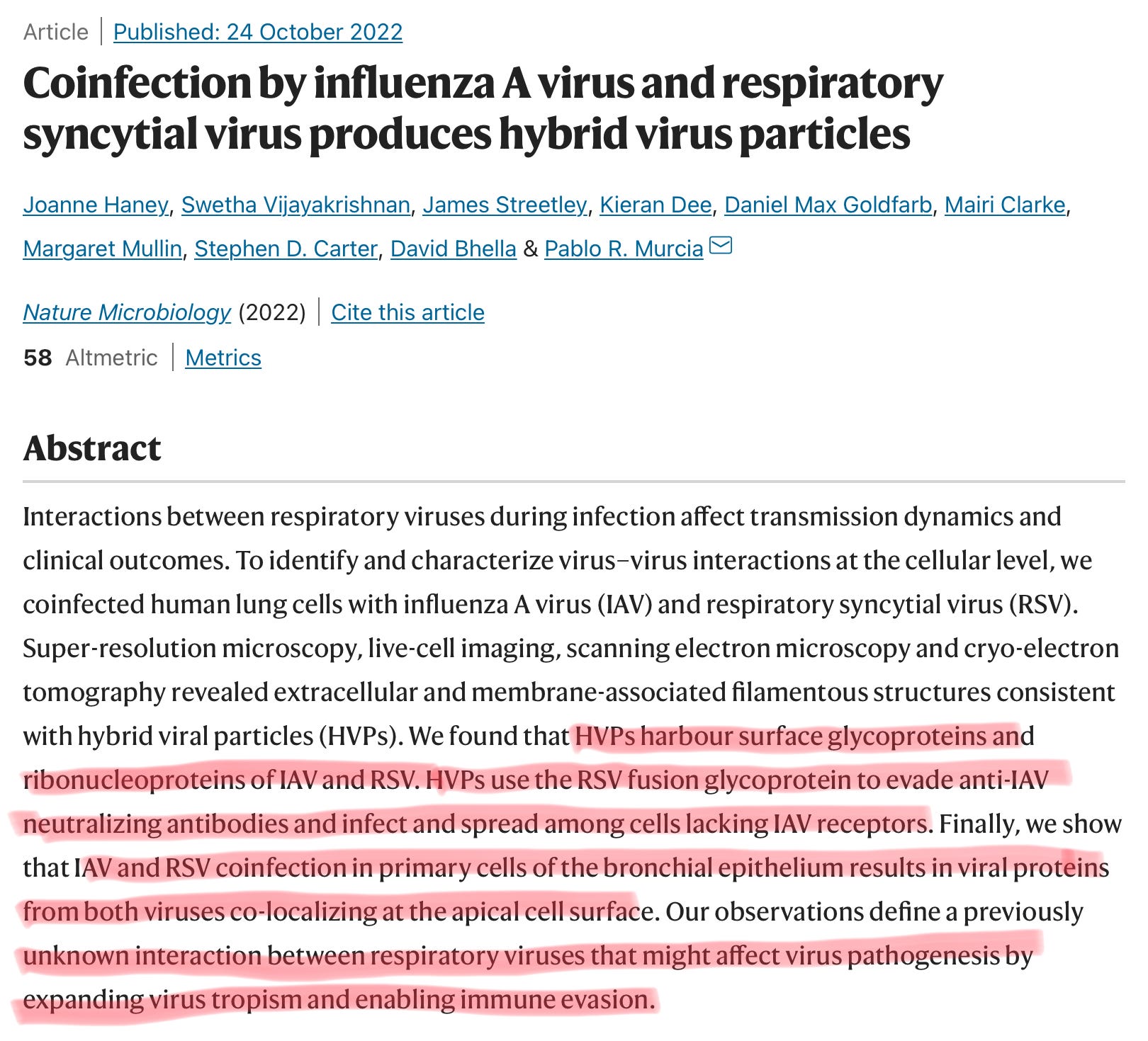
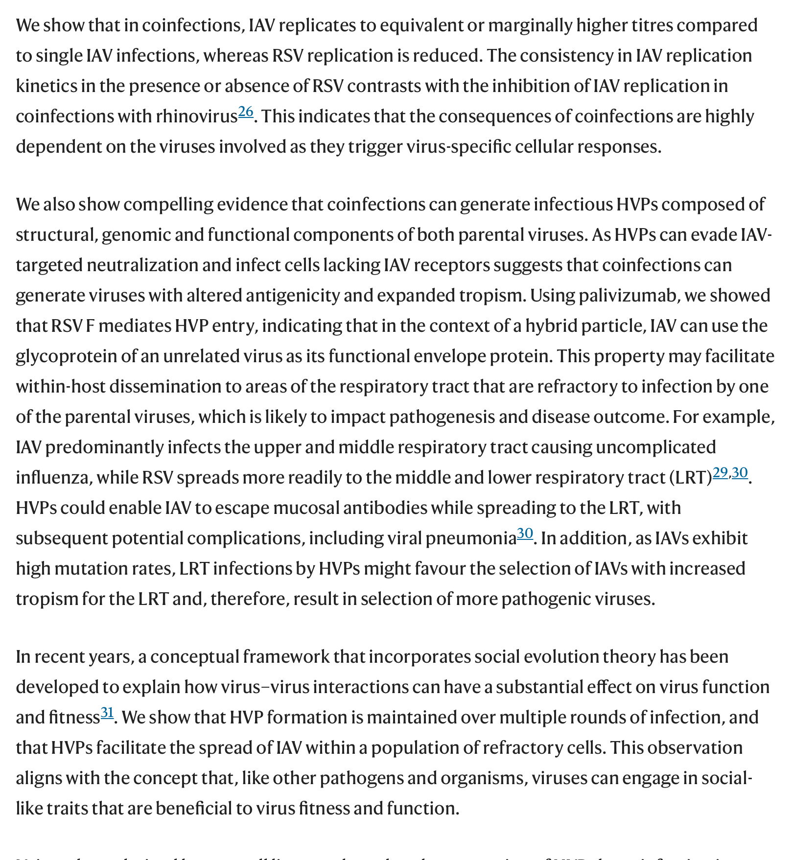
Co-infections with virus and even different pathogens is not something new, in truth, this has been one of the most observed phenomena since the start of the pandemic, where one common trend among severe cases of SARS-CoV-2 was often the co-infection or subsequent infection with bacteria, which certain bacteria being present at much higher levels than others (Prevotella being one of those). But viruses creating hybrid particles is something novel, where these particles contain the genetic information and key parts of both viruses.
But the really mind-blowing part of these hybrid particles can “exchange” parts, in which one of these particles could contain Influenza A infecting a new cell using the entry mechanism of RSV, this is a very powerful method of immune evasion.
This was never one of my usual insane observations, but it falls in line with one I will propose at the end of this.
Viruses Can Scatter Their Genes Among Cells and Reassemble
Not only are some viruses split into multiple segments that infect host cells separately, but as researchers in France have now discovered, those fractured viruses can flourish with their genomes scattered like puzzle pieces across a multitude of host cells. Something — presumably, the diffusion of molecules among the infected cells — allows complete viral particles to replicate, self-assemble and infect anew.
“You can get all of the necessary gene products together to produce new viruses in a cell that doesn’t actually have all the gene segments in it,” explained Christopher Brooke, a virologist at the University of Illinois at Urbana-Champaign.
“A classical view in virology assumes that the viral replication cycle occurs within individual cells,” said Anne Sicard, the lead author of the new study and a plant pathologist at the French National Institute for Agricultural Research (Institut National de la Recherche Agronomique, or INRA) in Montpellier. But in the case of this “multipartite” virus that she and her colleagues examined, “it seems that this is not true. The segments infect cells independently and accumulate independently in the plant host cells.” She added, “It really shows that the virus doesn’t work at a single-cell level, but at a multicellular level.”
Multipartite viruses have been known for over half a century, when researchers realized that a virus could be composed of two or more independent pieces, all of which were vital for infection. One piece might be necessary for making essential viral enzymes, for instance, while the other would be needed to make the capsule in which the viral particles (or virions) are packaged and transported to other cells.
Zwart points out that most ideas about the advantages of multipartition are really about genome segmentation, not partition of the virus into different infective units. Separating the genome into segments allows different viruses to recombine various advantageous forms of their genes easily.
Arvind Varsani, a virologist at Arizona State University, agrees. “From a modularity perspective, you could see the advantages of multicomponent viruses where each module can be independent,” he said. In a plant with multiple coinfecting viruses, “you can gain elements much more quickly, and adapt to the environment by mixing and matching.”
Proof of the strength of this strategy can be found in the influenza virus, a master of reassortment. The flu genome also has eight segments, although those segments are packaged together in one viral capsule. That allows it to reap the benefits of segmentation without paying all the costs of multipartition.
Asking how viruses operate as populations rather than individual virion particles is going to end up being important for lots of different viral systems.
I often find myself delving deeper into the fringe (as the original meaning of the world, cutting edge) science, the “non-canonical” side because it often bears more fruits for my questions and pursuits than conventional paper mill-esque science.
While this was done in plants, given the first paper covered here, it is not too stretched of the imagination that this is happening in other species too, especially in humans. SARS-like viruses are well known to be prone to recombination inside hosts, recombining to such a degree they can achieve molecular exchange, changing only just one amino acid. I believe many viruses are able to scatter their genes as a means to “exchange upgrades”.
Leading us to one of my favorite articles of the last few years, the internet of viruses and bacteria.
‘Broadband’ Networks of Viruses May Help Bacteria Evolve Faster
A newly discovered mechanism may enable viruses to shuttle genes between bacteria 1,000 times as often as was thought — making them a major force in those cells’ evolution.

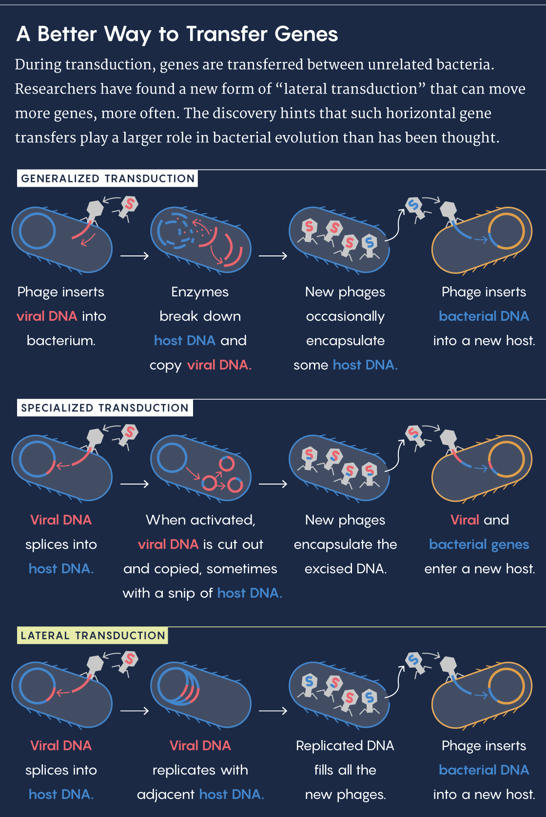
Bacteria have a sneaky evolutionary advantage: their own version of the internet for swapping survival solutions. It’s a living network of viruses that can shuttle genetic information between unrelated cells. Known as transduction, this process is one of the ways that bacteria can bypass the generation-by-generation plodding of vertical inheritance and instead share information horizontally, enabling genes slowly shaped by natural selection to enter a new population in an instant.
Scientists have known this transduction network must influence bacterial evolution across the sweep of centuries, but they presumed its short-term impact might be limited because transduction events seemed rare. A study published last week in Science, however, discovered a new mechanism of transduction that occurs 1,000 times more often, and that may accelerate bacterial evolution to a similar degree. Transduction may in fact be a central force in bacterial evolution.
“We had assumed that transduction occurred at rates analogous to dial-up internet,” said John Chen, an assistant professor of microbiology and immunology at the National University of Singapore and a lead co-author on this study. “But it appears that in some cases, transduction rates are more akin to broadband.” Their results not only suggest that transduction makes horizontal gene transfer much more common, but also that this bacterial internet might have been shaped by selection for the benefit of both bacteria and the viruses called bacteriophages.
A bacteriophage has another option, too: Its genes can slip into the bacterium’s genome unnoticed and lie in wait. Known as a prophage, this stretch of foreign DNA can persist for generations before activating. Approximately half of all sequenced bacteria contain a prophage, and many house more than one.
Once activated, the prophage cuts itself out of the bacterial genome, replicates and then packages its DNA into phages. Occasionally, there’s an error in the cutting, and a small portion of the bacterial genome gets copied and packaged alongside the prophage. When that bacteriophage infects another host, the bit of bacterial DNA gets incorporated into the new host’s genome in a process called specialized transduction.
Another mystery that lateral transduction could solve is the existence of “pathogenicity islands” — chunks of bacterial genomes that contain a genetic toolkit for evading antibiotic resistance and becoming more virulent. They dot the genomes of many pathogenic species of Staphylococcus, but are oddly absent in their nonpathogenic close relatives. Scientists struggled to explain this pattern, but lateral transduction provides a plausible mechanism.
For the reader unaware, I have for long (since circa mid-2020) argued that SARS-CoV-2 either had a bacteriophage phase, or it was a bacteriophage, and ever since some research was published arguing the same, while not strictly a bacteriophage itself, it certainly behaves like one.
In my pursuit of making sense of what was going on “under the hood”, and to explain why and how there was so much bacterial involvement within SARS-CoV-2 while researching I had many ideas. One of these was if the Spike Protein could interact with LPS, confirmed by recent research, another one is a mixture of my and a friend’s observations.
Given that the Spike Protein interacts with LPS via lipid-lipid interactions, among other mechanisms, it is now safe to propose that the Spike will also interact with bacteria, it explains a lot of the shift in quantities of specific bacteria in moderate to severe cases, why some guts and microbiomes are impacted so heavily, the translocation of said bacteria. And since this explains so much of what is going on with bacteria and the virus-bacteria interaction, it might explain one of our points so far “unexplored”. Biofilms.
Here is one Substack I wrote talking about the potential of SARS-CoV-2 and biofilm interaction. It is clear that the virus will do what is described at the end of that post, and more, since the Spike Protein can interact with bacteria, and it will play a role in biofilms, it wouldn’t be a surprise if under certain conditions the describe above occurs in stringent conditions, often akin to immune suppression, or inflammatory dysregulation.
Only time and research will tell, but now I am confident enough to write this, not sound completely crazy, one could argue if in the near future some variant of Omicron acquires a mutation present in Alpha, or unusual mutations not seen in over a year. I highly recommend you to read the entire paper referred to in the biofilm substack above “biofilms as a potential reservoir”.
From my perspective, it would make much more sense for SARS-CoV-2 to create a reservoir inside other places, rather than our cells, so far most evidence points out that SARS-CoV-2 infection of our cells inevitably leads to the death of that cell.
Addition prior to publishing:
Mere minutes after publishing this, I stumbled upon this paper.
SARS-CoV-2 Spike Protein and Mouse Coronavirus Inhibit Biofilm Formation by Streptococcus pneumoniae and Staphylococcus aureus
The presence of co-infections or superinfections with bacterial pathogens in COVID-19 patients is associated with poor outcomes, including increased morbidity and mortality. We hypothesized that SARS-CoV-2 and its components interact with the biofilms generated by commensal bacteria, which may contribute to co-infections. This study employed crystal violet staining and particle-tracking microrheology to characterize the formation of biofilms by Streptococcus pneumoniae and Staphylococcus aureus that commonly cause secondary bacterial pneumonia. Microrheology analyses suggested that these biofilms were inhomogeneous soft solids, consistent with their dynamic characteristics. Biofilm formation by both bacteria was significantly inhibited by co-incubation with recombinant SARS-CoV-2 spike S1 subunit and both S1 + S2 subunits, but not with S2 extracellular domain nor nucleocapsid protein. Addition of spike S1 and S2 antibodies to spike protein could partially restore bacterial biofilm production. Furthermore, biofilm formation in vitro was also compromised by live murine hepatitis virus, a related beta-coronavirus. Supporting data from LC-MS-based proteomics of spike–biofilm interactions revealed differential expression of proteins involved in quorum sensing and biofilm maturation, such as the AI-2E family transporter and LuxS, a key enzyme for AI-2 biosynthesis. Our findings suggest that these opportunistic pathogens may egress from biofilms to resume a more virulent planktonic lifestyle during coronavirus infections. The dispersion of pathogens from biofilms may culminate in potentially severe secondary infections with poor prognosis. Further detailed investigations are warranted to establish bacterial biofilms as risk factors for secondary pneumonia in COVID-19 patients
The detection of the SARS-CoV-2 spike protein after bacterial co-incubation (Figure 6) suggests that the interaction between spike protein and S. pneumoniae likely involved direct adhesion of the S1 subunit to bacterial surface molecule(s).
From the results of this in vitro study, we propose that SARS-CoV-2 and related coronavirus infections may trigger an active dispersion of bacteria from biofilm.
I have no snark remarks to be made, in the fact most of our ideas and hypothesis being proven right in the last 8 weeks. Now it is not a matter of if, but how, when, and where.
The Spike Protein disrupts the biofilms and displaces the LPS from bacteria to its surface.

The paper above directly correlates with the Endotoxin one… I wish you all a happy Sunday.
A big thank you to the supporters here and on Kofi, and people who share posts they find interesting.
Excellent work! Yes, we are in the middle of an ‘experiment in nature’ where we are the subjects (guinea pigs). It seems to me that the mechanisms you describe here could all fall under the heading of “Unintended Consequences of Unleashing Untested Vaccines.”
Thank you so much for compiling and sharing such fascinating information!!!
Just yesterday, I learned about common building blocks of SARS with other viruses on the absolutely wonderful mejbcart Substack cited below:
https://mejbcart.substack.com/p/how-much-hiv-is-in-sars-cov-2-faucis
"....In order to propagate for example viral SARS-CoV-2 infection with a big pool of identical building blocks common with HIV-1, with Marburg, with all the other viruses mentioned in previous posts, incredible large portions of HUMAN proteins, any genetic re-programming of the human body for synthetic SARS Spikes production will automatically affect infections with other even more lethal or maybe even benign viruses."
On to bacteria....
And, now, I am so happy to be able to share your Substack research on bacteria with two io groups.
Carlo Brogna and his team have another cool study. https://f1000research.com/articles/11-292
We know that the virus can enter bacteria and stick around for a long period. Further, a most intriguing finding, Different lineages? Hijacking of the cellular machinery of two different species? "Overall, this finding provides an indication that bacteria might be a potential source of novel SARS mutations, and gives rise to the possibility that intra-host SARS haploytpes might reflect different intestinal bacterial prior host environments of the virus. This observation would represent the basis for one of the proposed origins of the Omicron variant i.e. that Omicron (and other variants) might have evolved in the gut bacteria of one person."